
There Is a Light Switch in the Brain
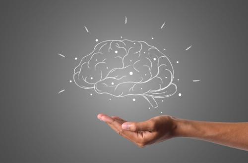
On 5 October 2026, the Nobel Assembly at the Karolinska Institutet in Stockholm awarded the 2026 Nobel Prize in Physiology or Medicine to three scientists from two countries: Peter Hegemann and Georg Nagel of Germany, and Karl Deisseroth of the United States. Hegemann is a professor at Humboldt University of Berlin, Nagel at the University of Würzburg, and Deisseroth at Stanford University and a researcher at the Howard Hughes Medical Institute. They were honoured for discovering light-gated ion channels and developing optogenetics.
Hegemann and Nagel discovered channelrhodopsin, a light-sensitive protein in single-celled green algae. Deisseroth then turned the protein into a light switch for nerve cells. The result is a method that allows neuronal activity in a living brain to be switched on or off with millisecond precision. The Nobel Committee described the discovery as the foundation for a new era in neuroscience.
The story behind the award began in a freshwater pond, home to the single-celled alga Chlamydomonas reinhardtii. It has neither eyes nor a brain, yet it always knows where to swim: towards the light. The seemingly simple question of how a cell without eyes can “see” led to Stockholm two decades later. Hegemann has acknowledged that, at first, almost no one was interested in or took seriously their research into green algae. That is the first lesson of this year’s Nobel: major revolutions often arise from small curiosities with no promised practical benefit.
The answer to the algae mystery turned out to be a protein. In 2002, Nagel and Hegemann’s team reported channelrhodopsin-1 (ChR1). When paired with retinal, a form of vitamin A also used by our eyes, it becomes a channel that allows protons to flow when exposed to light. A year later, they discovered its counterpart, channelrhodopsin-2 (ChR2), which proved much more useful. ChR2 opens very quickly after absorbing a photon and allows various cations, both monovalent and divalent, to pass through. What makes it special is that this single protein performs two tasks at once: it detects light and acts as an ion channel. In rhodopsin in vertebrate eyes, these functions are separated by a long chain of biochemical reactions.
The decisive step came from Stanford in 2005. Deisseroth’s team introduced the ChR2 gene into mammalian neurons using a lentivirus as a vehicle, then illuminated them with flashes of blue light. The result was reliable control over each electrical burst of a neuron on a millisecond scale, including excitatory and inhibitory synaptic transmission. For the first time, researchers could “type” patterns of electrical activity into neurons with a precision matching the brain’s own language. The technique became known as optogenetics.
What was new about it? Before optogenetics, neuroscience had two main tools for influencing the brain, and both were crude. Electrical electrodes worked quickly but stimulated all nearby cells indiscriminately. Drugs could be more selective chemically, but acted slowly, over minutes or hours.
Optogenetics combines three properties that had never before been brought together. First, it is genetically specific, allowing only particular cell types to be fitted with the switch. Second, it offers temporal precision measured in milliseconds. Third, it requires only a single protein component. Other protein variants, such as halorhodopsin, can even switch neurons off using a different colour of light. There is now both an “on” switch and an “off” switch.
Its greatest paradigm shift is epistemological. For decades, neuroscience was largely a science of correlation. fMRI and EEG tell us which areas “light up” when someone is afraid or falls in love. But activity occurring at the same time does not mean that one thing causes the other. Optogenetics allows neuroscience to ask two of the most rigorous causal questions: is a group of neurons necessary for a behaviour, which can be tested by switching it off? And is it sufficient to produce that behaviour, which can be tested by switching it on? Neuroscience has moved from observing the brain to interrogating it.
One of the most dramatic examples comes from memory research. In 2012, Susumu Tonegawa’s team at MIT, working with Deisseroth, marked neurons in the dentate gyrus of mice’s hippocampi that were active when the mice learned to fear. Those neurons were fitted with ChR2. When the researchers illuminated them in a different environment, the mice immediately froze in fear, while control mice did not. The conclusion was clear: activating a small, specific group of neurons within a memory engram was enough to recall that memory. The idea that memories are stored in concrete physical traces, once speculative, now had experimental evidence.
In understanding disease, optogenetics acts like a conceptual surgical tool. Deep brain stimulation (DBS) has long been used to treat Parkinson’s disease, but its mechanism has remained unclear because electrodes affect a mixture of cell types. In 2009, Deisseroth’s team used optogenetics to systematically switch different circuit components on and off in freely moving mouse models of Parkinson’s disease. They found that the therapeutic effect in the subthalamic nucleus could be explained by direct stimulation of incoming afferent axons.
This means a therapy used in patients for years was found to work through a pathway different from the one originally assumed. The same logic is now being applied to depression, addiction, anxiety, epilepsy and chronic pain. Brain disorders are increasingly understood as circuit disorders, rather than simply imbalances in chemical substances.
What about humans? The first clinical step came in the eye, an organ that naturally receives light. In 2021, Sahel, Roska and colleagues reported on a patient with retinitis pigmentosa who had been blind for decades. The patient was injected with an AAV viral vector carrying the light-sensitive protein ChrimsonR, and then wore special light-stimulating goggles. After treatment, the patient was able to see, locate, count and touch objects using the treated eye—something that had been impossible before the injection.
The strength of this approach is that it does not depend on the particular mutation. Hundreds of different genetic mutations can cause retinitis pigmentosa. Optogenetics does not correct the mutation; instead, it gives surviving retinal cells a new ability to detect light.
Even so, we must be honest about its limitations. Almost all optogenetics findings still come from animal models, and applying the technique to the human brain faces three major obstacles. First, it requires gene therapy, with all its risks, including immune responses to foreign proteins from algae. Second, light has difficulty penetrating deep brain tissue without implanted optical fibres. Third, mouse circuits are not always identical to human circuits. Ethical questions are also only beginning to be discussed.
If memories can be recalled—and, in animals, even created artificially—where is the boundary between treatment and the manipulation of identity? The history of this discovery also reminds us that science is a collective endeavour. Ernst Bamberg, Edward Boyden and Gero Miesenböck also helped pioneer the field, but Nobel rules limit the number of recipients to three.
Ultimately, the 2026 Nobel is a story about how fundamental knowledge that appears “useless” can become the key to a deeper understanding of ourselves. A protein that helps algae swim towards the sun is now helping us understand how memories are stored, how fear is evoked and how blind people may see again.
To me, its deepest meaning is this: thoughts, memories and feelings have biological addresses that can be traced, tested and perhaps one day repaired. But precisely because we now have the switch, a question older than neuroscience has become more urgent: who has the right to touch it, and for what purpose?