
Indian Scientists Create World's Most Detailed 3D Digital Map of the Human Brain
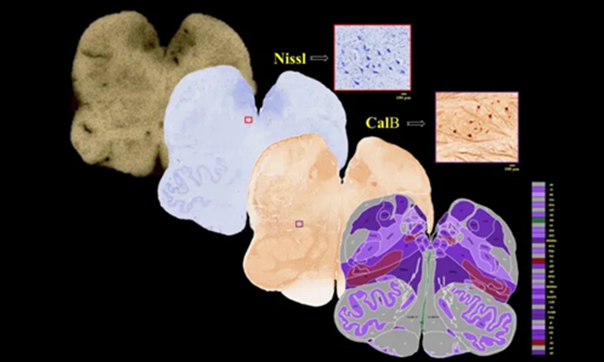
For more than a century, scientists have studied the human brain much like early cartographers mapping unknown territory, piecing together a broad landscape from scattered observations. To bridge this vast gap, researchers at the Sudha Gopalakrishnan Brain Centre (SGBC) at the Indian Institute of Technology Madras (IIT-M) have created the world’s most detailed three-dimensional (3D) digital atlas of the human brainstem, with cellular-level resolution.
The digital map, named Anchor (Atlas of Neurochemical Characterisation of the Human Brainstem with 3D Reconstruction), combines more than 500 tissue slices from the brains of foetuses, children and adults. Using high-resolution microscopic imaging technology, the project identified more than 200 brain cell clusters and neural pathways without the need for expensive molecular techniques.
The brainstem is a vital part connecting the brain to the spinal cord, controlling crucial functions such as breathing, heart rate, sleep and movement. Its extremely dense architecture has long made this region difficult to map in detail.
The scientific project has received high praise from the international community for successfully integrating MRI scans showing the whole brain with cellular pathology that views the brain cell by cell.
“We are looking at a visionary programme that places India at the international table,” said Shubha Tole, an Indian neuroscience expert from the Tata Institute of Fundamental Research, who described the project as an “unprecedented integration” of engineering, neuroscience and medicine.
Until now, pathologists diagnosing neurological disorders such as Alzheimer’s have generally examined only a dozen or so tissue samples from an organ that actually contains some 86 billion neurons. As a result, most of the brain’s landscape has remained unseen.
“As a neuropathologist, I begin by examining the whole brain with the naked eye before looking at small pieces under the microscope,” said Rebecca Folkerth, an expert affiliated with Harvard Medical School and New York University who collaborated on the project.
“For Alzheimer’s disease, we may examine only 15 to 20 sections, a tiny fraction of one per cent of the entire organ.”
Folkerth added that this digital map fulfils her long-standing dream of being able to match brain scans with the brain’s microscopic anatomy. Through Anchor, users can now accurately zoom in from the external structure of the brainstem on an MRI down to the level of individual neurons.
Although not a direct diagnostic tool, the atlas, which is freely available online, has broad potential medical applications. By comparing healthy and diseased tissue, scientists can deepen their understanding of Parkinson’s disease, stroke, Alzheimer’s, autism, and even the long-term effects of infections such as Covid-19.
Partha Mitra, a brain scientist at Cold Spring Harbor Laboratory in New York involved in the research, said that a detailed brain atlas such as this could have a “transformative impact” on the study of neurological diseases.
Looking ahead, the SGBC now plans to map more than 100 whole human brains across a range of ages and neurological conditions in order to create an increasingly comprehensive digital reference library.